Details of the molecular structure of key brain proteins revealed by breakthrough Alzheimer’s research give new insights for potential targeted therapies.
Scientists researching Alzheimer’s have described for the first time the structure of molecules within a human brain. Details published in Nature today describe how the researchers used cryo-electron tomography, bolstered by fluorescence microscopy, to probe the depths of a brain donated by an Alzheimer’s patient.
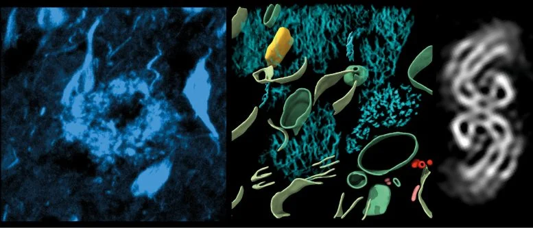
This provided 3-dimensional maps, in which it became possible to visualize proteins, the molecular building blocks of life a million times smaller than a grain of rice, inside a brain. In this study, focusing on two proteins that cause dementia, zooming in became possible.

Left, fluorescence image of amyloid in cryo-preserved post-mortem human brain. Middle, 3-dimensional molecular architecture of β-amyloid plaque. Right, in-tissue structure of tau filaments within post-mortem brain. Credit: University of Leeds
This study identified the molecular structure of tau in tissue, how the amyloids are arranged, and new molecular structures entangled within this pathology in the brain. Dementia represents the highest cause of natural death in the UK; Alzheimer’s is the most common one.
Impact of Molecular Structures on Alzheimer’s
Indeed, both β-amyloid plaques and abnormal tau filaments of Alzheimer’s disease are thought to disrupt cell-cell communication, causing memory loss, confusion, and cell death.
Dr Rene Frank, Associate Professor in the University of Leeds’s School of Biology and lead author, said: “This first glimpse at the molecular structure of molecules inside the human brain offers further clues as to what happens to proteins in Alzheimer’s disease but also sets out an experimental approach that can be applied to better understand a broad range of other devastating neurological diseases.”.
Over the course of more than 70 years, thousands of scientists in laboratories around the world, each doing biochemistry on proteins one at a time in a test tube, have achieved an enormous catalog of molecular structures. It had also become clear that most functions in biology were a product of an orchestra made up of many different proteins.
This research, led by the University of Leeds working in collaboration with scientists at Amsterdam UMC, Zeiss Microscopy, and the University of Cambridge, represents just one of the many exciting new areas in which structural biologists are working directly inside cells and tissues—in situ—to examine how proteins function fitted together and on one another in human cells and tissues ravaged by disease. This observation of the interplay of proteins within tissues, during a longer period of time, will give impetus to the identification of new targets for the next generation of mechanism-based therapeutics and diagnostics.
This breakthrough Alzheimer’s research not only uncovers the molecular structure of some key brain proteins in detail but also is an important step toward unlocking the detailed mechanisms behind the disease itself. The molecular composition of β-amyloid plaques and tau filaments in the human brain will be determined with an unprecedented level of detail. This new knowledge will be integral in developing targeted therapies to disrupt or mitigate the detrimental impact on brain function caused by the activity or presence of these proteins in the processes leading to Alzheimer’s pathology.
This study has made use of cryo-electron tomography and fluorescence microscopy to map β-amyloid and tau 3-dimensional structures at unprecedented resolution. The approach followed in this research—an examination of the dynamics of proteins within their native tissue environment rather than in isolation—presents a more realistic representation of interactions and other behaviors of proteins in the complex biological context of the Alzheimer’s disease-affected brain.
According to Dr. Rene Frank, a lead author of the study from the University of Leeds, there is a wider significance of this research beyond Alzheimer’s alone. It will be pioneering techniques to study molecular structures within intact tissues that will let researchers begin to unpack the complexity of neurological disorders related to protein misfolding and aggregation. This new approach thus holds out hope for new avenues toward an understanding of the mechanisms behind diseases and new therapeutic strategies.
This is an example of one such interdisciplinary effort by researchers at the University of Leeds, Amsterdam UMC, Zeiss Microscopy, and the University of Cambridge, to firmly fix what are complex biomedical challenges. These scientists, through their experience in structural biology, microscopy techniques, and neurosciences, challenge the boundaries of what can be imagined in understanding the molecular basis for Alzheimer’s disease and related disorders.
Looking ahead, the detailed molecular insights gained in this work will inform future studies aimed at identifying and validating new therapeutic targets. Treatments targeting specific molecular pathways implicated in Alzheimer’s disease are in development, with interventions early in the disease process, slowing or probably even halting progression. This constitutes a ray of hope for millions who suffer from the symptomatic holocaust of Alzheimer’s and their families and is illustrative of the potential of basic science for revolutionizing global health challenges.
Second, in this work, cryo-electron tomography together with fluorescence microscopy has been used for the first time in structural biology, which, by any standard of the imagination, is an enormous feat in technological advancement. These are techniques that offer researchers excellent views of molecular structures, unrivaled to date, in a very dynamic way of structure behaviors in their native environment. Scientists can, therefore, examine β-amyloid plaques and tau filaments within intact brain tissue to study their spatial distribution and interactions at an unprecedented level of detail. It is for this reason that this holistic approach to the intricate game of different proteins playing various roles makes sense in neurodegenerative diseases, Alzheimer’s included.
This study at the University of Leeds and its collaborators exemplifies another step toward making biomedical research more integrative and translational. Scientists make a linkage between basic science discoveries and clinical applications to fast-track effective diagnostics and treatments for Alzheimer’s disease. This gives more information on the molecular underpinnings of the illness and attempts to underline the importance of cooperation across many disciplines in treading multifaceted health challenges.
In doing so, this study provided results that are very promising in laying a foundation for future work while reminding us that relentless pursuit of innovation and exploration in neuroscience and structural biology is demanded. Elucidating the role proteins play in Alzheimer’s disease pathology is researchers’ forging a pathway toward personalized medicine approaches that one day will revolutionize neurological disorder diagnosis and treatment. It is hoped that these efforts will finally improve the outcomes and quality of life of those affected, aligning with evolving scientific understanding and improving technology in the cases of AD and related diseases.
Reference: “CryoET of β-amyloid and tau within postmortem Alzheimer’s disease brain” by Madeleine A. G. Gilbert, Nayab Fatima, Joshua Jenkins, Thomas J. O’Sullivan, Andreas Schertel, Yehuda Halfon, Martin Wilkinson, Tjado H. J. Morrema, Mirjam Geibel, Randy J. Read, Neil A. Ranson, Sheena E. Radford, Jeroen J. M. Hoozemans and René A. W. Frank, 10 July 2024, Nature.
